Knowing how dental anatomy works is essential for good oral health. Whether you live in Holt or are just wondering what your teeth are made of, this walking guide will help you understand how detailed your dental anatomy is and how each part does its job with the help of Holt dentist.
The Structure of a Tooth
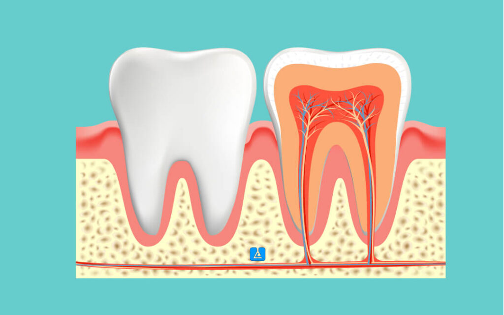
Your tooth is made up of several different layers, all of which have a different role to play. Here’s a deeper dive into these layers:
- Enamel: Enamel is the top layer covering the tooth and the top delicate tissue in the human body. Protects the inner layers of the tooth from decay and trauma. Enamel consists primarily of hydroxyapatite, a crystal form of calcium phosphate.
- Dentin: Dentin is a porous yellowish tissue that lies beneath the enamel. It constitutes the bulk of the tooth. Dentin supports the enamel and transmits sensory signals through the outer layers of the tooth to the nerve in the pulp. It is less hard than enamel and contains tiny tubules that traffic through nutrients and sensations.
- Pulp: Pulp refers to the innermost tooth consisting of nerves, blood vessels, and connective tissues. It nourishes the tooth and transmits signals, like pain, to the brain. The pulp stretches from the crown to the ends of the roots.
- Cementum: A hardened layer surrounding the roots of teeth. By connecting via the periodontal ligament, the root keeps the tooth anchored in the jawbone. Cementum is less hard than either enamel or dentin but is important for keeping teeth stable.
- Periodontal Ligament: It refers to a well-fitted, specialized group of connective tissue fibers that connect the tooth to the alveolar bone socket. Absorbs the shock of chewing, and helps to hold the tooth in place.
Humans have four different types of teeth, each adapted for different functions:
- Incisors: The four upper and four lower front teeth. Sharp, chisel-shaped teeth are used for cutting food into smaller, more easily swallowed pieces.
- Canines: There are four canines, one next to each of the incisors. Canine teeth are pointed and are used to tear and grasp food. They also assist in guiding teeth into place as the mouth closes.
- Premolars (Bicuspids): Eight premolars, four on each side of the mouth, are located behind the canines. The flat surfaces with ridges of a premolar’s crown crush and grind food.
- Molars: The largest teeth, molars are twelve in total, including the wisdom teeth. Molars possess wide, flat surfaces for crushing food into smaller pieces for swallowing, suitable for later digestion.
Dental Numbering Systems
Dentists have a couple of systems for identifying and referring to teeth:
- Universal Numbering System: It designates every tooth a number starting with the upper right third molar (tooth #1) around the arch to the upper left third molar (tooth #16) then the lower left third molar (tooth #17) around to the lower right third molar (tooth #32).
- International Numbering System (FDI): This system of FDI (Foreign Direct Investment) is common and widely used outside the US. It uses a two-digit number for each tooth, the first digit representing the quadrant, and the second digit representing the tooth’s position in that quadrant.
Conclusion
The significance of preserving good oral health is highlighted by the fascinating and intricate topic of dental anatomy. You can better appreciate the importance of consistent dental care if you are aware of the anatomy and function of your teeth. Do not be afraid to speak with a local dentist in Holt if you are worried about the state of your teeth. They can offer professional guidance and care.


